How do health care professionals diagnose the problem?
On initial assessment at Next Step Physio your physiotherapist will perform an examination that will start with a thorough history. They will ask questions about when the pain began, when and where precisely the pain occurs, your activity levels, whether you have had any previous spinal pain or problems in the past, if you have had previous treatment, what makes your pain better or worse, whether there are any noted muscle weaknesses or tingling sensations, and will ask about whether you have any problems with urination or bowel movements (particularly incontinence). They will also want to know if you have pain in any other areas of your body such as your hips, knees, or shoulders. They may also ask questions about your sport, school, or work activities.
A physical examination will be done once the history is complete.
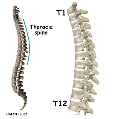
Your physiotherapist will examine your thoracic spine along with the other areas of the back to evaluate the curves of the spine, spasm of the muscles, unusual markings on the skin or soft tissues along the spine and will assess your overall posture and alignment of the back as well as your lower extremities. Your physiotherapist will palpate, or touch along the spine and over the muscles to determine if any particular areas are painful or tight. They may push on the spine, or manually move the spine to get a general idea of how much motion is available at each segment.
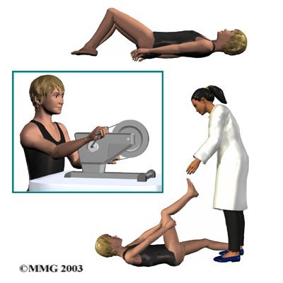
Your physiotherapist will also examine your hips, knees, and ankles to determine if these joints and the muscles that are involved with them might be contributing to the pain you feel in your back. The length (flexibility) and strength of the muscles of the buttocks, the front of the hip, as well as the thigh (quadriceps and hamstrings muscles) are particularly important areas that your physiotherapist will assess. These muscles can create an abnormal pull on the back if they are too tight, or not support the back well enough if they are too weak. Both tightness and/or weakness can contribute to your back pain. The hip joints themselves, if restricted in their ability to move through a full range of motion, can particularly contribute to back pain so their motion will be thoroughly assessed.
Your physiotherapist will also want to examine your ability to bend your back forwards, backwards, sideways, as well as rotate it and to get into positions involving a combination of these motions. Assessing this movement in the thoracic spine may include having you raise your arms up overhead or put them behind your neck.
Your physiotherapist will also look at your posture and alignment while you are standing and sitting, and may also want to watch you during different activities such as walking, squatting, jumping, lifting one leg, or kneeling on your hands and knees. As your physiotherapist observes you performing these activities they will determine the ability for you to support your trunk with the deep muscles of the abdominal area and back.
A neurological examination may need to be done which will include checking your reflexes, sensation, and muscle strength.
After a thorough history and physical examination Scheuermann’s disease may be suspected. The only way to definitely diagnose Scheuermann's kyphosis, however, is with an X-ray.
Investigations
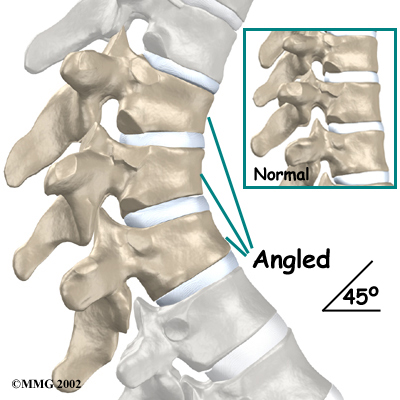
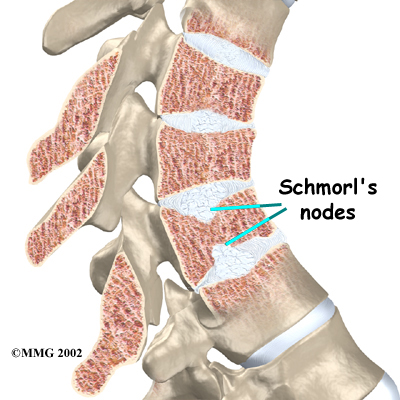
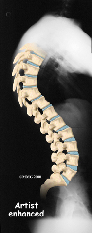
Taken from the side, an X-ray may show vertebral wedging, Schmorl's nodes, and changes in the vertebral end plates. Doctors use X-ray images to measure the angle of kyphosis. An official diagnose of Scheuermann's disease is made when three vertebrae in a row wedge five degrees or more and when the kyphosis angle is greater than 45 degrees.
A side-view X-ray can also show if the spine is flexible or rigid. Patients are asked to bend backwards and hold the position while an X-ray is taken. The spine straightens easily when it is flexible. In patients with Scheuermann's disease, however, the curve stays rigid and does not improve by trying to straighten up.
From the front, X-rays show if the spine curves from side to side. This sideways curve is called a scoliosis and occurs in about one-third of patients with Scheuermann's kyphosis.
X-rays can also show signs of wear and tear in adults (spondylolysis) who have extra lumbar lordosis from years of untreated Scheuermann's disease.
A Computed Tomography scan (CT) may be ordered. This is a detailed X-ray that lets doctors see slices of the body's tissue.
Myelography is a special kind of X-ray test. For this test, dye is injected into the space around the spinal canal. The dye shows up on an X-ray. This test is especially helpful if the doctor is concerned whether the spinal cord is being affected.
Magnetic resonance imaging (MRI) may be ordered. An MRI uses magnetic waves rather than X-rays to show the soft tissues of the body. This machine creates pictures that look like slices of the area being examined. This test does not require special dye or a needle.
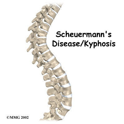
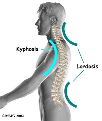
Scheuermann's disease or kyphosis, once definitively diagnosed, is determined as either being typical (Type I) or atypical (Type II). These two forms of the disease affect different parts of the spine. The typical form (most common type) has the thoracic kyphotic pattern described in this section. In these cases the lower (lumbar) spine compensates by developing an increased lordosis curve. This curve is termed ‘hyperlordotic’, which means the curve has increased beyond what is considered “normal.”
The atypical form of Scheuermann’s disease (Type II) affects the low back known as the lumbar spine. It is the upper part of the lumbar spine (where the thoracic spine transitions to become the lumbar spine) that is involved. Type II is seen most often in young boys before puberty who are active in sports activities. They experience pain that goes away with rest and changes in position or activity level.
Next Step Physio provides services for physiotherapy in Edmonton.
