Open surgical treatment for spinal compression fractures due to osteoporosis is rarely needed. (Open procedures require larger incisions to give the surgeon more room to operate.) In rare cases of severe trauma, however, open surgery is sometimes required. Open surgery is done if the spinal segment has loosened and bone fragments have damaged the spinal cord and spinal nerves.
Surgeons have begun using two new procedures to treat compression fractures caused by osteoporosis. Both are considered minimally invasive. Minimally invasive means the incisions used are very small, and there is little disturbance of the muscles and bones where the procedure is done. These two procedures help the fracture heal and avoid the problems associated with more involved surgeries.
These new procedures are:
- vertebroplasty
- kyphoplasty
Vertebroplasty
This procedure is most helpful for reducing pain. It also strengthens the fractured bone, enabling patients to rehabilitate faster.
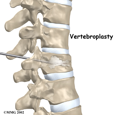
To perform vertebroplasty, the surgeon uses a fluoroscope to guide a needle into the fractured vertebral body. A fluoroscope is a special X-ray television that allows the surgeon to see your spine and the needle as it moves. Once the surgeon is sure the needle is in the right place, bone cement, called polymethylmethacrylate (PMMA), is injected through the needle into the fractured vertebra. A reaction in the cement causes it to harden within 15 minutes. This fixes the bone so that it does not collapse any further as it heals. More than 80 percent of patients get immediate pain relief with this procedure.
Next Step Physio's Guide to Vertebroplasty
Kyphoplasty
Kyphoplasty is another way for surgeons to treat vertebral compression fractures. Like vertebroplasty, this procedure halts severe pain and strengthens the fractured bone. However, it also gives the advantage of improving some or all of the lost height in the vertebral body, helping prevent kyphosis.
Two long needles are inserted through the sides of the spinal column into the fractured vertebral body. These needles guide the surgeon while drilling two holes into the vertebral body. The surgeon uses a fluoroscope (mentioned above) to make sure the needles and drill holes are placed in the right spot.
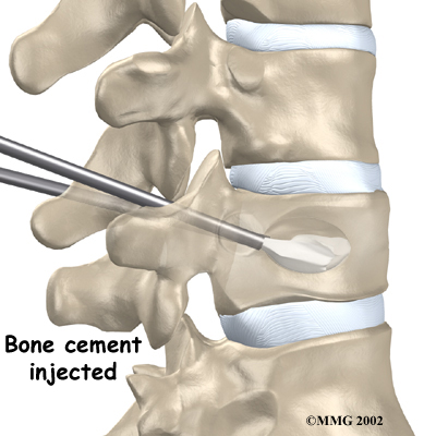
The surgeon then slides a hollow tube with a deflated balloon on the end through each drill hole. Inflating the balloons restores the height of the vertebral body and corrects the kyphosis deformity. Before the procedure is complete, the surgeon injects bone cement into the hollow space formed by the balloon. This fixes the bone in its corrected size and position.
A Patient's Guide to Kyphoplasty
Portions of this document copyright MMG, LLC.
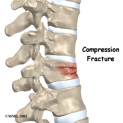


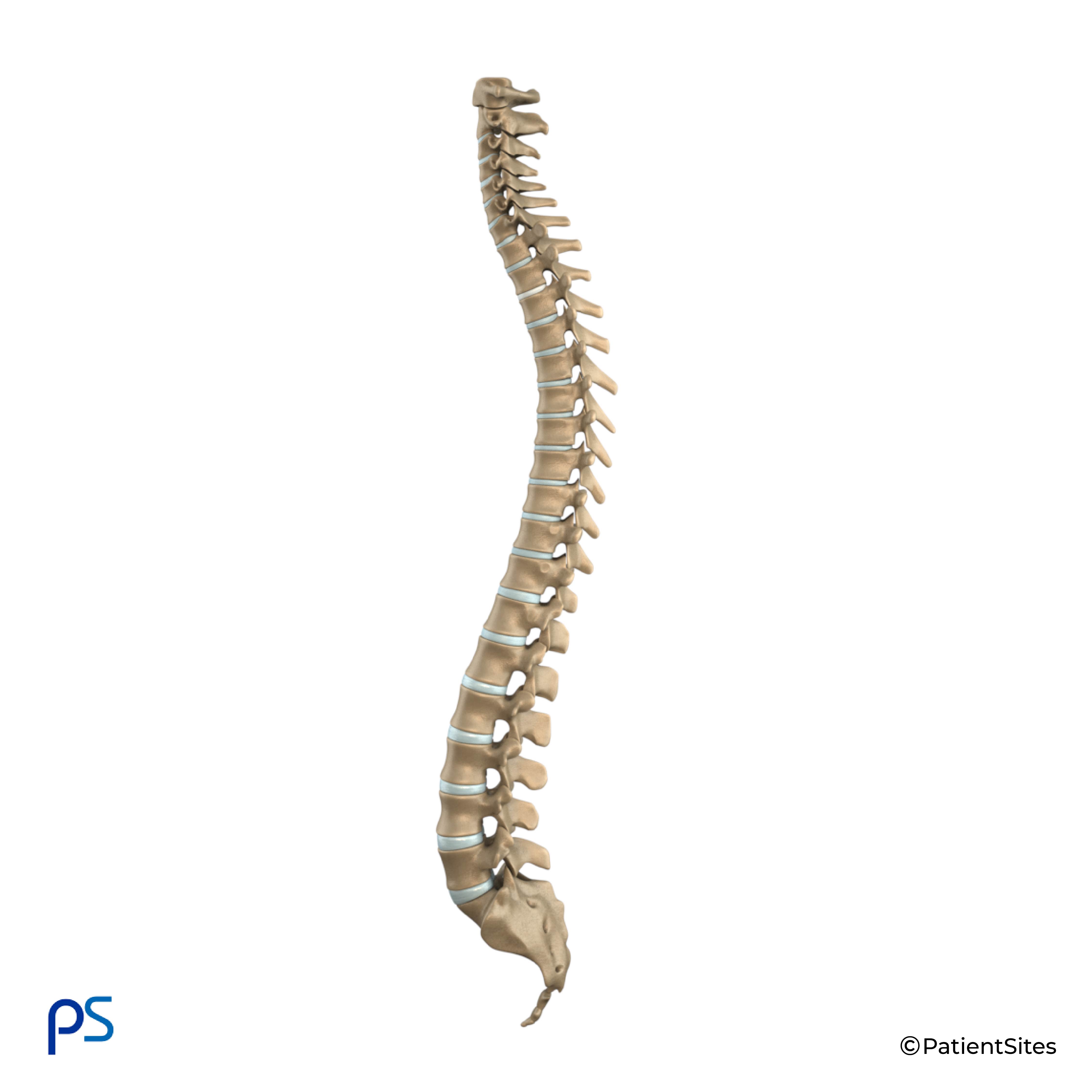
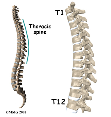
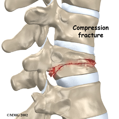
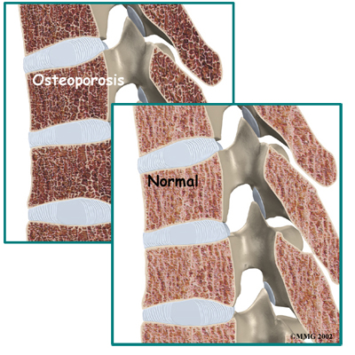 Strong, healthy bones are able to withstand the forces and strains of normal activity. Compression fractures in the spine happen when either the forces are too great or the bones of the spine aren't strong enough. The vertebral body cracks under pressure. Fractures from forceful impact on the spine tend to crack the back (posterior) part of the vertebral body. Fractures from osteoporosis usually occur in the front (anterior) part of the vertebral body.
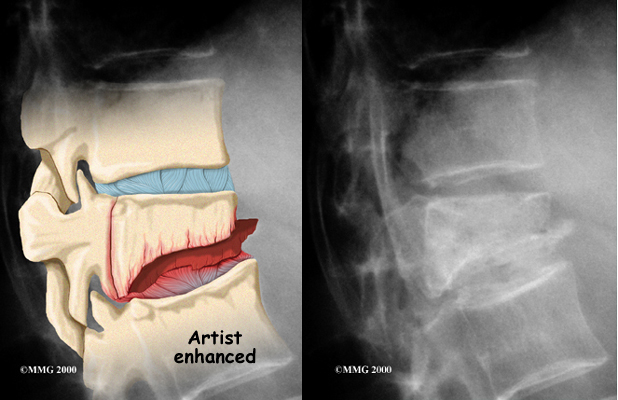 If your doctor believes there is a compression fracture,
 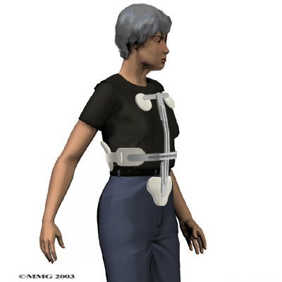 The majority of patients with compression fractures are treated without surgery. Most compression fractures heal within eight weeks with simple remedies of medicine, rest, rehabilitation, and a special back brace.
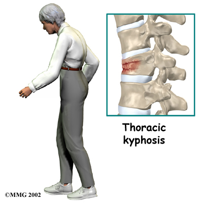
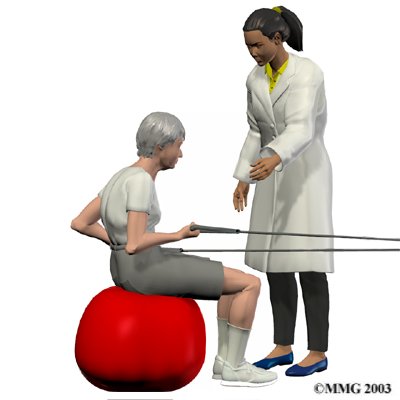 Your physiotherapist will prescribe exercises for you to do in the clinic and also to be done as part of a home program. Exercises that improve the range of motion in your back, neck, shoulders as well as your hips may be prescribed. If your compression fracture was from osteoporosis, then the extension motion of your upper back (thoracic spine) will be of paramount importance. As mentioned above, wedge compression fractures of the thoracic spine from osteoporosis often lead to a flexed back posture. The risk of losing the ability to function in the upright extended position is high so maintaining this motion is crucial. Even the proper use of your shoulder joints will suffer if the spine loses extension therefore exercises may also be prescribed to maintain shoulder function. Neck range of motion can also be affected if the flexed posturing becomes severe thus range of motion exercises for the neck may also be required. Hip range of motion deficits will be addressed as normal hip range of motion allows the spine to move more freely and decreases the stress on the spinal joints. Patients with traumatic stress fractures don’t often present with the wedge shaped fractures and therefore the primary focus will be the recovery of all ranges of motion, not just thoracic extension.
